Echocardiography
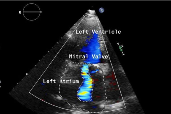
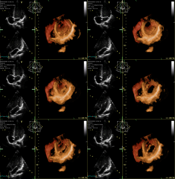
Echocardiography, also called an echo test or heart ultrasound, is a test that takes “moving pictures” of the heart with sound waves. Echocardiography is a simple and painless procedure that uses sound waves to take “pictures” of your heart.
Why do you need an echo test?
Your doctor may use an echo test to look at your heart’s structure and check how well your heart is working.
This test may be needed if…
- You have a heart
- You’ve had a heart
- You have unexplained chest
- You’ve had rheumatic
- You have a congenital heart
How is it done?
Echo tests are done by trained sonographers. You’ll lie on a bed on your left side or. The sonographer will put special jelly on a probe and move it over your chest. Ultra-high-frequency sound waves will pick up images of your heart and No X-rays will be used. Your heart’s movements can be seen on a monitor. It’s painless and has no side.
What can the test show?
- The size and shape of your heart

- How well your heart is working overall
- If a wall or section of heart muscle is weak and not working correctly
- If you have problems with your heart’s valves
- If you have a blood clot
What happens after the echo?
- If the test results show some problems you’ll be advised to see your cardiologist.
There is a highly qualified English-speaking cardiologist at Agape Medical Center – Dr. Larisa Pavlova who is trained to do the echo test and can also give you a consultation and prescribe a treatment if needed.
